


组织透明化
您现在的位置 > 产品中心
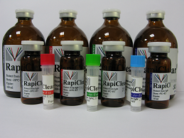
RapiClear 1.47组织透明试剂
▪ 样品可以直接从水,缓冲溶液和甘油转移到RapiClear 1.47透明剂中。
1. Grundy L et al. Chronic linaclotide treatment reduces colitis-induced neuroplasticity and reverses persistent bladder dysfunction. JCI Insight (2018).https://doi.org/10.1172/jci.insight.121841
Drosophila:
1. Benavides LR et al. Phylogeny, evolution and systematic revision of the mite harvestman family Neogoveidae (Opiliones Cyphophthalmi). Invertebrate Systematics (2019). https://doi.org/10.1071/IS18018
6. Liu N et al. Functional variants in TBX2 are associated with a syndromic cardiovascular and skeletal developmental disorder. Hum Mol Genet (2018).https://doi.org/10.1093/hmg/ddy146
产品介绍:
RapiClear 1.47
品牌:SunJin Lab
▪ 不同于脂溶性透明,此透明剂为水溶性透明剂,使用方便、透明可逆。
▪ 样品可以直接从水,缓冲溶液和甘油转移到RapiClear 1.47透明剂中。
▪ 如果样品重新浸入水或缓冲溶液中,透明效果是可逆的。
▪ RapiClear 1.47即用型,无需离心。
▪ RapiClear 1.47使得组织表面以下0.5 mm处目标清晰可见。
▪ RapiClear 1.47的应用不会引入样品变形。
产品规格:
RC147001 10mL
RC147002 100mL
产品应用:
▪ 动物组织切片~0.5mm厚度
▪ 生物材料
▪ 昆虫
▪ 植物
▪ 斑马鱼幼鱼
相关产品:
▪ iSpacer imaging Spacer/成像垫片(货号# IS001)
▪ RapiClear CS mounting solution(货号# RCCS001)
▪RapiClear CS mounting gel(货号# RCCS004)
相关文献:Mouse:
1. Grundy L et al. Chronic linaclotide treatment reduces colitis-induced neuroplasticity and reverses persistent bladder dysfunction. JCI Insight (2018).https://doi.org/10.1172/jci.insight.121841
2. Atlan G et al. The Claustrum Supports Resilience to Distraction. Curr Biol(2018). https://doi.org/10.1016/j.cub.2018.06.068
3. Baranska A et al. Unveiling skin macrophage dynamics explains both tattoo persistence and strenuous removal. J Exp Med (2018).https://doi.org/10.1084/jem.20171608
4. Mondor I et al. Clonal Proliferation and Stochastic Pruning Orchestrate Lymph Node Vasculature Remodeling. Immunity (2016).http://dx.doi.org/10.1016/j.immuni.2016.09.017
5. Seiradake E et al. FLRT structure: balancing repulsion and cell adhesion in cortical and vascular development. Neuron (2014).http://dx.doi.org/10.1016/j.neuron.2014.10.008
Drosophila:
1. Benavides LR et al. Phylogeny, evolution and systematic revision of the mite harvestman family Neogoveidae (Opiliones Cyphophthalmi). Invertebrate Systematics (2019). https://doi.org/10.1071/IS18018
2. Göpel T et al. Morphological description, character conceptualization and the reconstruction of ancestral states exemplified by the evolution of arthropod hearts. PLoS One (2018). https://doi.org/10.1371/journal.pone.0201702
3. Marcogliese PC et al. IRF2BPL Is Associated with Neurological Phenotypes. Am J Hum Genet (2018). https://doi.org/10.1016/j.ajhg.2018.07.006
4. Lin G et al. Phospholipase PLA2G6, a Parkinsonism-Associated Gene, Affects Vps26 and Vps35, Retromer Function, and Ceramide Levels, Similar to α-Synuclein Gain. Cell Metab (2018). https://doi.org/10.1016/j.cmet.2018.05.019
5. Li-Kroeger D et al. An expanded toolkit for gene tagging based on MiMIC and scarless CRISPR tagging in Drosophila. eLife (2018).https://doi.org/10.7554/eLife.38709.001
6. Liu N et al. Functional variants in TBX2 are associated with a syndromic cardiovascular and skeletal developmental disorder. Hum Mol Genet (2018).https://doi.org/10.1093/hmg/ddy146
7. Lee PT et al. A gene-specific T2A-GAL4 library for Drosophila. eLife (2018).https://doi.org/10.7554/eLife.35574
8. Lee PT et al. A kinase-dependent feedforward loop affects CREBB stability and long term memory formation. eLife (2018).
https://doi.org/10.7554/eLife.33007.001
9. Myers L et al. The Drosophila Ret gene functions in the stomatogastric nervous system with the Maverick TGFβ ligand and the Gfrl co-receptor. Development.(2018). http://dx.doi.org/10.1242/dev.157446
10. Frank DD et al. Early Integration of Temperature and Humidity Stimuli in the Drosophila Brain. Curr Biol (2017). http://dx.doi.org/10.1016/j.cub.2017.06.077
11. Enjin A et al. Humidity Sensing in Drosophila. Curr Biol (2017). http://dx.doi.org/10.1016/j.cub.2016.03.049
12. Osterfield M et al. Diversity of epithelial morphogenesis during eggshell formation in drosophilids. Development (2015).
http://dev.biologists.org/lookup/doi/10.1242/dev.119404
13. Nagarkar-Jaiswal S et al. A library of MiMICs allows tagging of genes and reversible spatial and temporal knockdown of proteins in Drosophila. eLife(2015). http://dx.doi.org/10.7554/eLife.05338
Porcine:
1. Yang, H., Yu, P.K., Cringle, S.J., Sun, X.*, Yu, D.Y.* (2015). Quantitative study of the microvasculature and its endothelial cells in the porcine iris. Exp Eye Res. 132: 249-258. doi: http://dx.doi.org/10.1016/j.exer.2015.02.006*corresponding authors
2. Yang, H., Yu, P.K., Cringle, S.J., Sun, X.*, Yu, D.Y.* (2015). Intracellular cytoskeleton and junction proteins of endothelial cells in the porcine iris microvasculature. Exp Eye Res. 140: 106-116. doi: http://dx.doi.org/10.1016/j.exer.2015.08.025 *corresponding authors
Zebrafish:
1. Steventon, B.*, Duarte, F., Lagadec, R., Mazan, S., Nicolas, J.F., Hirsinger, E. (2016). Species-specific contribution of volumetric growth and tissue convergence to posterior body elongation in vertebrates. Development. 143: 1732-1741. http://dev.biologists.org/lookup/doi/10.1242/dev.126375 *corresponding author
Human:
1. Yang, H., Yu, P.K., Cringle, S.J., Sun, X., Yu, D.Y.* (2017). Microvascular Network and Its Endothelial Cells in the Human Iris. Curr Eye Res. 43(1):67-76. doi: https://doi.org/10.1080/02713683.2017.1379544 *corresponding author